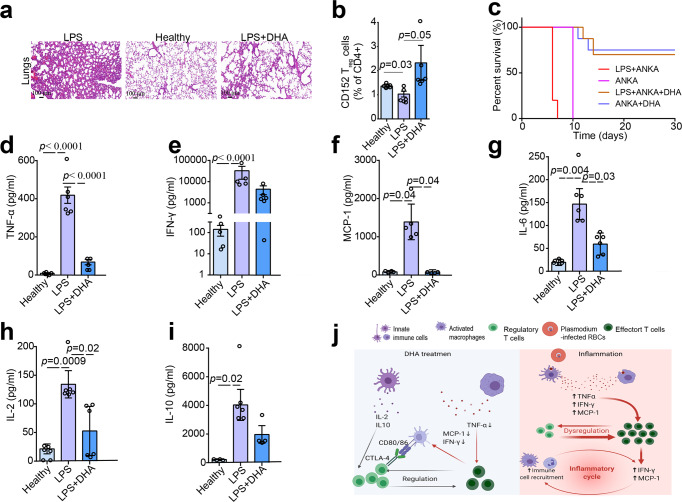
Fig. 3. Dihydroartemisinin (DHA) alleviated the inflammation in mice induced by LPS and P. berghei infection.
a Representative sections of HE stained lung tissues of mice treated by LPS, DHA. LPS treatment increased the number of inflammatory cells in the alveolar spaces, destructed the pulmonary interstitium, and ruptured collagen fibers compared to that from the control group. DHA mitigated LPS-induced acute lung injury in mice. b Bar graphs represent significant decrease in percentages of Treg in LPS treated group compared to healthy group, while significant increase in percentages of Treg in DHA treated group compared to LPS treated group. Proportion of total CD152+ Treg gated off the total CD4+ populations (n = 6, two-tailed Student t-test). c Survival curve of mice following single P. berghei ANKA infection or combined with LPS treatment under DHA treatment or without DHA treatment. The P. berghei ANKA-infected mice treated with LPS survived four days less than those only infected with P. berghei ANKA. On the contrary, DHA prolonged the survival time of P. berghei ANKA-infected mice treated with LPS (n = 8–10). Mice injected with LPS were previously infected with P. berghei ANKA for 4 days in LPS + ANKA group. d–i Serum abundance of IFN-γ, tumor necrosis factor α (TNFα), IL-2, MCP-1, IL-6, and IL-10 increased in LPS-treated mice compared with healthy mice while significantly decreased in DHA-treated mice (n = 4–6, two-tailed Student t-test). The sera were collected at day 7 after DHA treatment and the levels of the cytokines were determined by cytometric bead array (CBA). j A schematic diagram of DHA in its modulation on immune cells. DHA inhibited LPS- and P. berghei ANKA- induced inflammation. Figure was created using BioRender and Agreement number was GC24OV5V07. The error bars indicate standard error.

