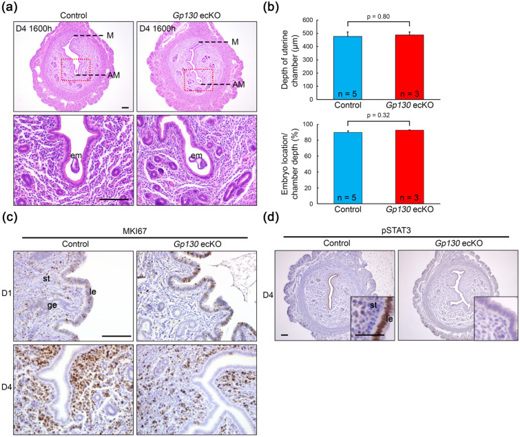
Figure 3.
Histological and histochemical characterization in Gp130 ecKO uterus before embryo implantation. (a) Representative images of uterine cross section of pre-implantation site on D4 (1600 h) from control and Gp130 ecKO. The depth of the uterine chamber (M-AM distance) is comparable between control and Gp130 ecKO (b, upper panel), and embryos are located in the deep antimesometrial side of the uterine chamber in both groups (b, lower panel). (c) Cell proliferation status detected by immunostaining of MKI67 at D1 (1000 h) and D4 (1600 h). (d) Immunostaining of phosphorylated STAT3 (pSTAT3) in pre-implantation uterus (D4, 1600 h). pSTAT3 is not detectable in the uterine epithelium from Gp130 ecKO. Scale bar, 100 µm (a,c, and d with lower magnification) and 50 µm (d with higher magnification). le luminal epithelium, ge glandular epithelium, st stroma, em embryo, M mesometrial side, AM antimesometrial side.

