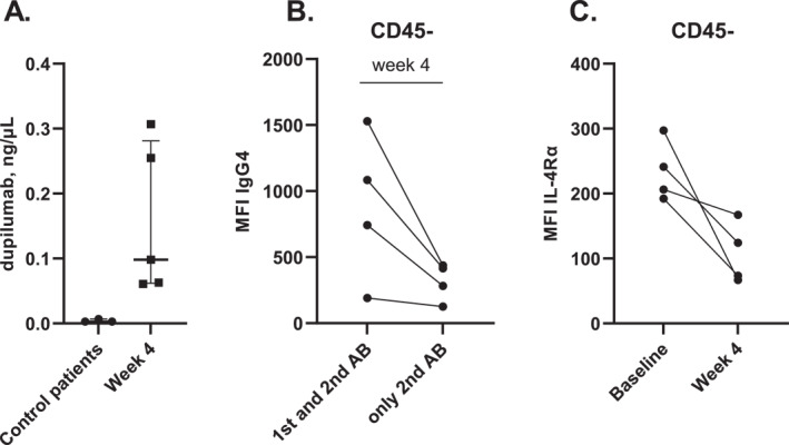
FIGURE 2.

Dupilumab measured in conjunctival cells obtained by conjunctival impression cytology. (A) Dupilumab measured in conjunctival cell suspensions from 5 dupilumab‐treated AD patients (at week 4) and 3 AD control patients (not treated with dupilumab) by using LC‐MS/MS. (B) Median Fluorescence Intensity (MFI) of IgG4 (= anti‐dupilumab) in CD45‐epithelial cells from 4 dupilumab‐treated AD patients (at week 4) compared to the control staining including only the secondary antibody (AB) streptavidine‐APC. (C) MFI of IL‐4Rα on CD45‐cells from 4 AD patients before dupilumab treatment (baseline) and after 4 weeks of treatment.
