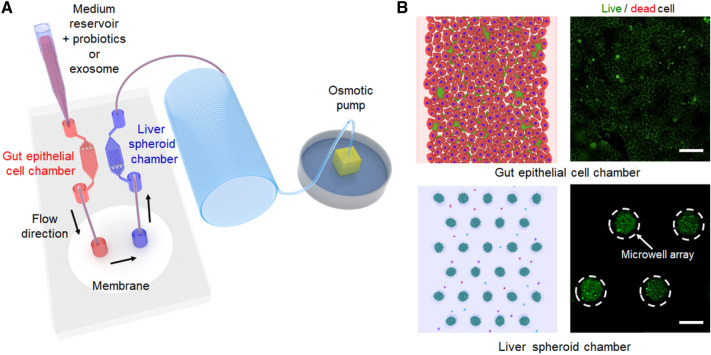
Fig. 1.
A The schematic of experimental setup of a gut-liver axis chip. The left chamber was used to establish an intestinal lumen using human epithelial Caco-2 cells. The right chamber, which contains microwell arrays, was employed to generate the 3D uniform-sized hepG2 spheroids. The continuous flow of the culture medium was introduced by an osmotic pump. B Schematic images of gut and liver spheroid chamber as well as representative immunofluorescence results showing that microbial and 3D hepG2 spheroids were located in the left gut chamber and right liver chamber of a gut-liver axis chip separated by cellulose membrane. Scale bar is 100 μm

