Figure 5.
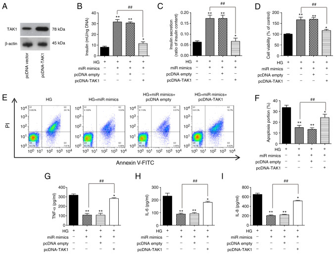
Overexpression of miR-143-3p attenuates HG-induced inflammatory response and apoptosis by targeting TAK1. (A) Western blotting was used to measure TAK1 expression after transient transfection with pcDNA-TAK1 in MIN6 cells. (B-I) MIN6 cells were co-transfected with the miR-143-3p mimics and pcDNA-TAK1 for 24 h, before they were cultured in 16.7 mM HG for an additional 24 h. (B) Insulin content and (C) insulin secretion were measured using an insulin ELISA kit. (D) Cell Counting Kit-8 assay was performed to detect cell viability. (E) Apoptosis was detected by flow cytometry and (F) was quantified. The levels of (G) TNF-α, (H) IL-6 and (I) IL-8 were measured using ELISA kits. Data are presented as the means ± SD from three individual experiments. *P<0.05 and **P<0.01 vs. HG; ##P<0.01 vs. HG + miR mimics group. miR, microRNA; TAK1, TGFβ-activated kinase 1; HG, high glucose.

