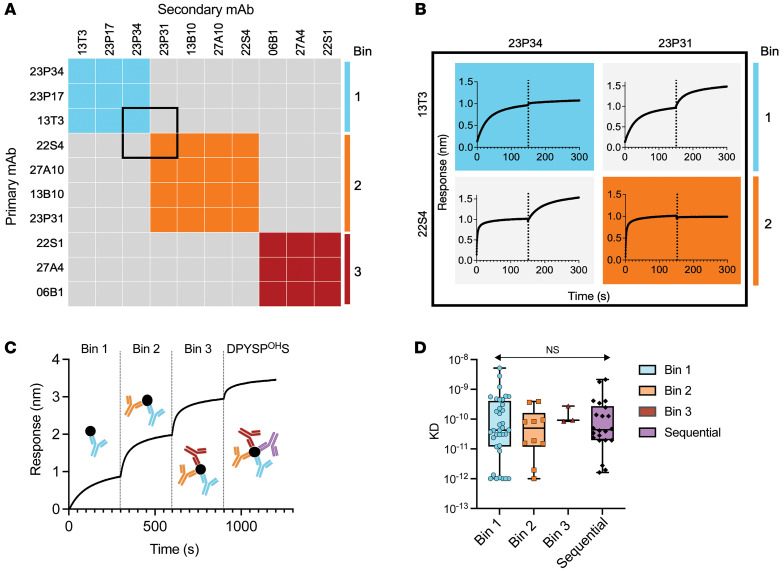
Figure 1. Conformational epitope binning of Ara h 2–specific antibodies.
(A) Representative epitope binning of nonsequential epitope Ara h 2 antibodies with an in-tandem epitope binning assay on BLI identified 3 conformational bins (colored boxes). The average response for binding of secondary binding antibodies was 0.77 ± 0.25 nm compared with 0.04 ± 0.05 nm for nonbinding secondary antibodies. (B) Inset with raw biolayer sensograms of the pairwise comparisons of primary (left) and secondary (top) Ara h 2–specific mAb binding to immobilized native Ara h 2. Primary and secondary antibody associations are divided by black dotted lines. (C) Sequential binding of epitope-specific antibodies to immobilized native Ara h 2. (D) Affinity of Ara h 2 antibodies for native Ara h 2 measured by BLI (shading by epitope). KD, dissociation constant. Box-and-whisker plots represent the mean, quartiles, and range. Statistical comparison was performed by 2-way ANOVA.

