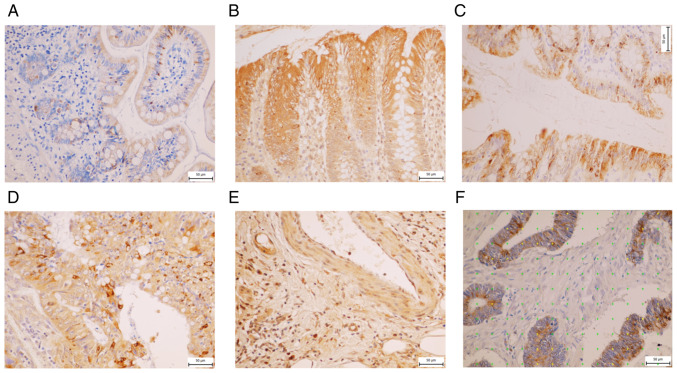
Figure 6.
Immunohistochemical staining of MUC13 in different parts of colon. (A) Non-malignant ileum, (B) non-malignant colon, (C) non-malignant rectum, (D) cytoplasm of cancer cells and (E) endothelial cancer cells. (F) Figure from Ellipse software of cancer cells using a point-counting method where yellow points stand for positivity for mucin 13, green points for negative.

