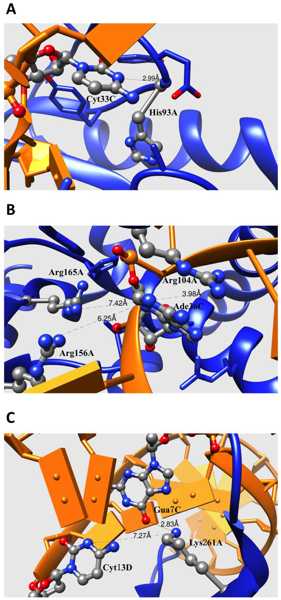
Figure 6:

(A) Fragment of binding interface of CCA-adding Enzyme complexed with tRNA (PDB: 3ovb). (B) Fragment of binding interface of TilS complexed with tRNA (PDB: 3a2k). (C) Fragment of binding interface of restriction endonuclease MspI on its palindromic DNA recognition site (PDB: 1sa3). The side chains of the residues directly contributing to the electrostatic interactions or H-bonding are shown with balls and sticks. The protein and DNA/RNA are marked as blue and orange. The distances between atom pairs are shown in Å.
