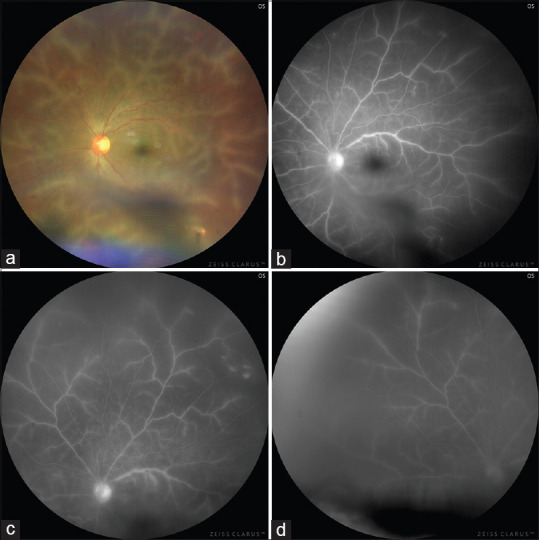
Figure 2.

Left eye on post-operative day 45. (a) Ultra-wide field image (Clarus 500, Zeiss, Carl Zeiss Meditech Inc., Dublin, USA) showing resolving vitreous hemorrhage, a normal optic disc, venous tortuosity, and translucent retinal perivascular infiltration affecting the venules from the posterior pole up to the periphery. (b-d) Fundus fluorescein angiography showing leakage of the affected vessels, late vessel staining, and peripheral capillary nonperfusion areas
