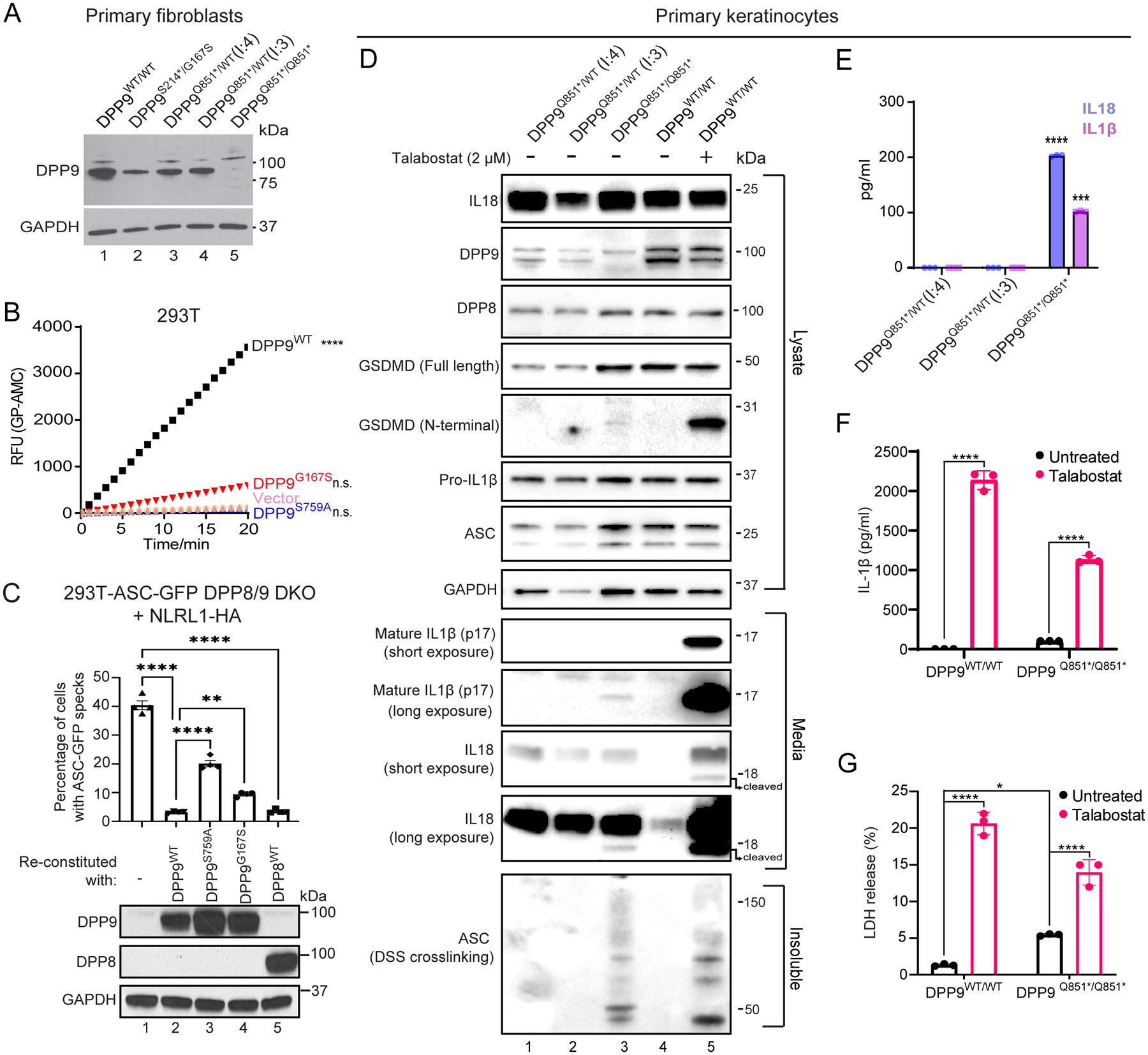
Fig. 2: Patients’ cells lack DPP9 or produce a strong enzymatic hypomorph.

(A) Western blot analysis of DPP9 in primary dermal fibroblasts of an unrelated control (WT/WT) (lane 1), affected individual from family 1 (proband (II:2) S214*/G167S) (lane 2), two unaffected individuals from family 2 ((I:4) and (I:3) Q851*/WT) (lanes 3 and 4 respectively) and affected individual from family 2 (proband (II:4) Q851*/Q851*) (lane 5).
(B) 293T cells were transfected with either vector, wild-type DPP9, DPP9 S759A or DPP9 G167S and lysed in PBS 1% Tween 20, 48 h after transfection. 0.3 μg of total lysate was then incubated with Gly-Pro-AMC fluorescence substrate. AMC fluorescence was measured for 20 mins at 25 °C in a 50 μl reaction every minute on a spectrometer. One-way ANOVA. ****P<0.0001 calculated by comparing the vector control to wild-type DPP9, DPP9S759A or DPP9G167S (n = 3 independent replicates).
(C). Top, quantification of ASC-GFP speck formation in DPP8/9 DKO 293T-ASC-GFP cells reconstituted with WT DPP9, DPP9 S759A, DPP9 G167S or WT DPP8, and transfected with NLRP1. The percentage of cells with ASC-GFP specks were counted in three different fields after fixation. More than 100 cells were scored per condition. Data are mean ± SEM, n = 4 independent replicates. **p < 0.01; ****p < 0.0001. Bottom, western blot analysis validating DPP9 and DPP8 expression.
(D) Western blot analysis of inflammasome components in primary keratinocytes of two unaffected individuals from family 2 ((I:4) and (I:3) Q851*/WT) and the affected individual from family 2 (proband (II:4) Q851*/Q851*). Additionally an unrelated control (+/+), treated with or without the potent DPP8/9 inhibitor Talabostat (2 μM) was included as a positive control for NLRP1 inflammasome activation.
(E) IL1β and IL18 ELISA of the culture supernatant from primary keratinocytes from the two unaffected individuals from family 2 ((I:4) and (I:3) Q851*/WT) and affected individual from family 2 (proband (II:4) Q851*/Q851*). ****p < 0.001, ***p < 0.01 (one-way ANOVA). (n=3 replicates).
(F), (G) Levels of IL-1β (F) and lactate dehydrogenase (LDH; G) released from DPP9WT/WT control and DPP9Q851*/Q851* patient-derived primary keratinocytes in absence or presence of Talabostat. Data are mean ± SEM, n = 3 independent replicates. *p < 0.05; ****p < 0.0001.
