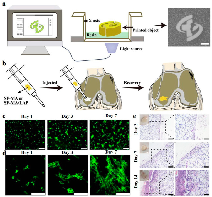
Figure 2.
3D Cryoprinting of LAP@SF injectable cryogels for bone tissue engineering. (a) Schematic illustration of the printing process of LAP@SF cryogels. (b) The printed cryogels can recover their original shape after injection and fit the tissue defect site. (c) Fluorescence staining image of live (green)/dead (red) BMSCs, after 1, 3, and 7 days of cultivation on the printed LAP@SF cryogels. Scale bar = 200 µm. (d) The morphology of BMSCs, after 1, 3, and 7 days of cultivation on the printed LAP@SF cryogels. Scale bar = 100 µm. (e) H&E staining of the printed LAP@SF cryogel scaffold after 3, 7, and 14 days of implantation; left, scale bar = 200 µm; right, scale bar = 100 µm. Reused with some rearrangement with permission of [74] (Under a Creative Commons license: CC BY-NC-ND 4.0).

