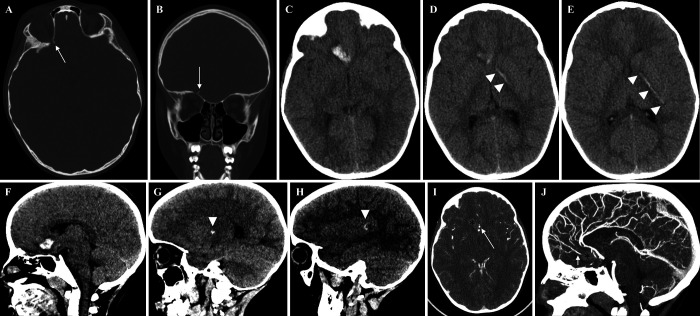
FIG. 2.
CT bone window showing a displaced fracture of the roof of the right orbit, with the fracture fragment (arrows) angulated superiorly toward the right basal frontal lobe (A and B). Axial (C–E) and sagittal (F–H) CT scans showing intraparenchymal hemorrhage in the right basal frontal lobe with linear extension (arrowheads) to the anterior limb of left internal capsule and left posterior insula. Findings suggest penetrating injury through the roof of the right orbit, extending across the right basal frontal lobe to the left internal capsule. In the contrast-supported CTA (I and J), the intraparenchymal hemorrhage (asterisks) is close to the ACAs (arrows). The intracranial branches of the internal carotid, basilar, and vertebral arteries are patent without signs of significant narrowing or intracranial dissection.

