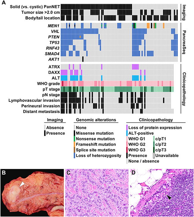
Figure 5.
(A) A summary of imaging findings, preoperative PancreaSeq testing, and postoperative clinicopathologic features of 87 PanNET patients. Both solid and cystic PanNETs exhibited similar genomic alterations; however, LOH for multiple genes correlated with several adverse clinicopathologic features, such as lymphovascular invasion, perineural invasion, higher T- and N-stage, distant metastases, loss of ATRX/DAXX expression, and the presence of ALT. (B) A representative example of a 1.5-cm PanNET (white arrowhead) in the pancreatic body that preoperative PancreaSeq testing revealed LOH for 4 genes. (C) Microscopically and immunohistochemically, the PanNET was classified as WHO grade 1. (D) However, within a single regional lymph node, a metastasis was identified (black arrowhead). In addition, the PanNET exhibited loss of ATRX expression and the presence of ALT. (C) Hematoxylin and eosin stain, magnification 400×. (D) Hematoxylin and eosin stain, magnification 200×.

