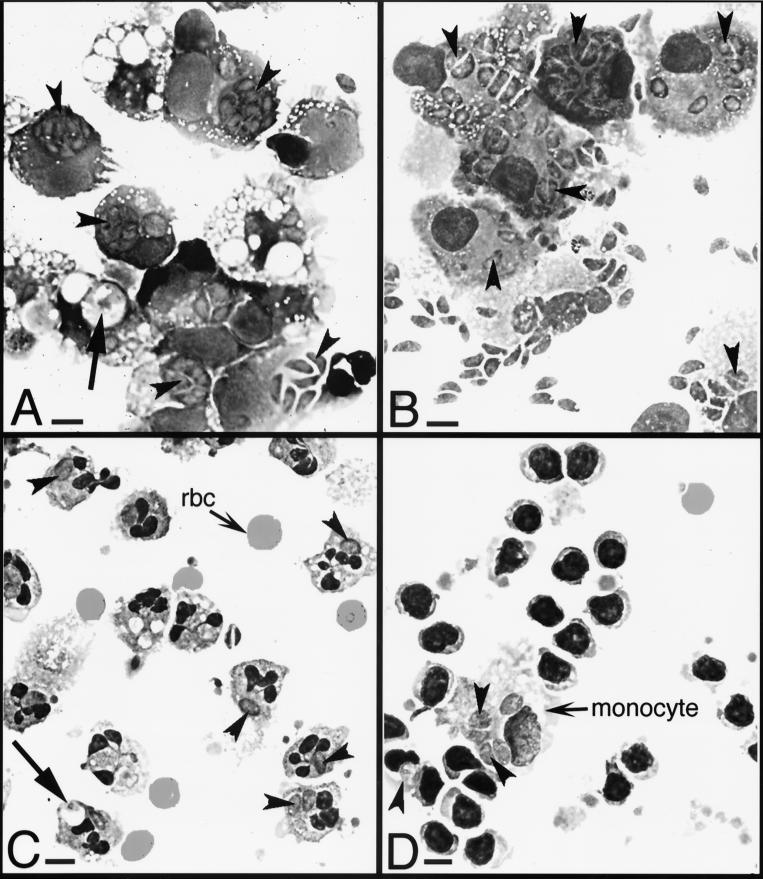
FIG. 2.
Photomicrographs of cytospin preparations of tachyzoite-infected host cells. Host cells were incubated for 24 h with tachyzoites at a cell-to-parasite ratio of 1:4. Arrowheads indicate parasites. Arrows indicate vacuoles containing digesting tachyzoites. Magnification, ×630. Bar = 2 μm. (A) Monocytes showing rapid tachyzoite division, as evidenced by up to eight tachyzoites per vacuole (see arrowheads). (B) Dendritic cells showing rapid tachyzoite division. (C) Neutrophils showing slow tachyzoite division, as seen by fewer than two tachyzoites per vacuole. rbc, red blood cells. (D) Lymphocytes showing slow tachyzoite division, even though tachyzoites in the contaminating monocyte were dividing rapidly. Photomicrographs were made using Kodak Elite chrome ASA 100 slide film, and 35-mm slides were scanned into Adobe Photoshop files using a SprintScan 35 (Polaroid Corp., Cambridge, Mass.).

