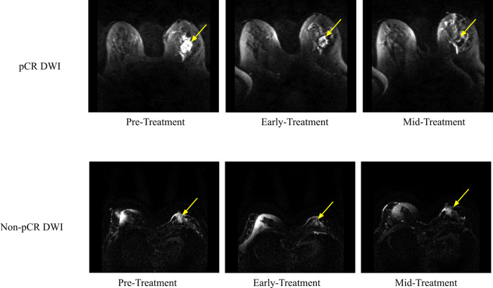
Fig 4.
DWIs for (A) a pCR patient and (B) a non-PCR patient at the pre-treatment, early treatment, and mid-treatment time points. For (A), the 54 year old Caucasian patient had invasive ductal carcinoma with HR- and HER2-. For (B), the 51 year old Caucasian patient had invasive ductal carcinoma with HR+ and negative HER2-. The yellow arrow indicates the approximate location of the tumor. DWI acquisitions were done using a b-value of 800 s/mm2. DWI: diffusion-weighted imaging).

