
Figure 1.
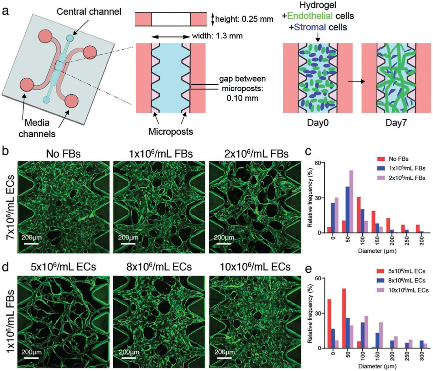
Seeding densities determine MVN diameters in microfludic devices. a) Schematic diagram of a microfluidic device with microposts for MVN formation. b) Representative images showing immortalized human umbilical vein EC (ImHUVEC, 7 × 10⁶ mL−1) MVNs made with lung FBs at 0, 1, or 2 × 10⁶ mL−1 concentration. ImHUVECs expressing blue fluorescent protein (BFP) were used. Green, ImHUVECs. c) Distribution of microvessel diameter of ImHUVEC MVNs made with or without FBs. Mean and SD of each group are: No FBs 164.3 ± 113.1 μm; 1 × 10⁶ mL−1 FBs 69.45 ± 60.78 μm; 2 × 10⁶ mL−1 FBs 47.97 ± 35.87 μm (n = 2 devices, 3 regions of interest (ROIs) each). Representative images (d) and diameter distribution (e) of MVNs made of ImHUVECs expressing BFP at 5, 8, or 10 × 106 mL−1 density with 1 × 10⁶ mL−1 lung FBs. Mean and S.D. of each group are: 5 × 10⁶ mL−1 ECs 36.28 ± 25.27 μm; 8 × 10⁶ mL−1 ECs 118.9 ± 93.68 μm; 10 × 106 mL−1 ECs 128.9 ± 79.19 μm (n = 2 devices, 3 ROIs each). Green, ImHUVECs.