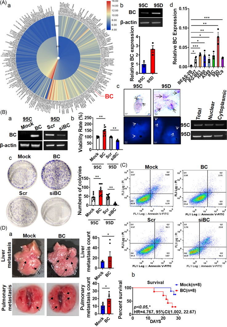
FIGURE 1.

BC overexpressed in lung adenocarcinoma (LUAD) cells, promoted lung cancer metastasis. (A) (a) Hierarchical clustering of 235 differentially expressed long non‐coding RNAs (lncRNAs) (|fold change| > 3) was performed using microarray data. Red, high expression; blue, low expression. (b) The expression of BC in 95C and 95D cells was analysed by RT‐PCR. (c) BC in nuclear and cytoplasmic fractions were detected by in situ hybridization, fluorescence in situ hybridization (left) and RT‐PCR (right). Arrows indicate positive hybridization sites. (d) BC expression was measured by RT‐qPCR in BEAS‐2B, PG‐LH7, PG‐BE1, H460, A549, PC9, 95C and 95D cells. (B) (a) Overexpression and knock‐down of BC were validated by RT‐PCR. (b) The viability of 95D cells was analysed by the MTT assay after BC overexpression or knock‐down. (c) Colony formation assays were performed with BC‐overexpressing and BC‐silenced cells. Quantitative analysis is shown on the right. (C) Flow cytometry was performed to measure cell cycle progression after ectopic expression or silencing of BC. (D) (a) Representative images of livers and lungs of nude mice, 4 weeks after tail‐vein injection (n = 8). Arrows indicate metastatic foci. Liver and pulmonary metastasis foci were quantitatively analysed. (b) The death rate of both groups is summarized. Data are represented as mean ± SEM of at least three independent experiments. *p < .05, **p < .01
