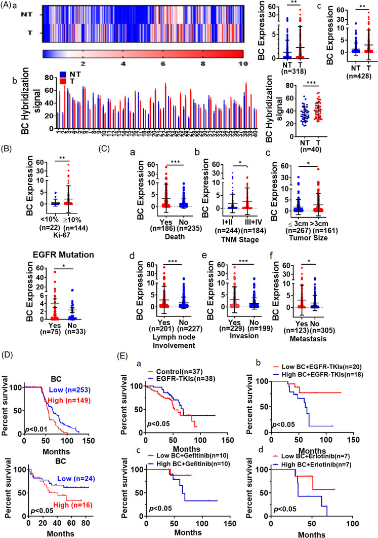
FIGURE 5.

BC was highly expressed in lung adenocarcinoma (LUAD) tissues. (A) (a) Differential BC expression between LUAD (T) tissues and adjacent non‐tumour (NT) tissues from 318 patients was determined by RT‐qPCR; (b) BC expression was measured by in situ hybridization in LUAD tissues and adjacent non‐tumour tissues from 40 patients; (c) BC expression analysis in the combined cohort (n = 428). (B) BC levels in LUAD tissues were compared between patients with different positive rate of Ki‐67 staining (upper). BC levels were measured in lung cancer tissues with and without EGFR mutation from 428 patients with LUAD (lower). (C) BC levels in LUAD tissues were compared between patients with different (a) survival, (b) TNM stage, (c) tumour size, (d) lymph node involvement, (e) invasion and (f) remote metastasis. Data are representative of at least three independent experiments. *p < .05, **p < .01. (D) Overall survival among 402 patients with LUAD was analysed according to BC expression levels measured by RT‐qPCR (upper). Kaplan–Meier analysis of 40 patients with LUAD showed differential survival corresponding to BC in situ hybridization signal levels (lower). (E) Kaplan–Meier analysis of overall survival of patients with LUAD that received EGFR‐tyrosine kinase inhibitor (EGFR‐TKI) treatment and conventional chemotherapy (a); Kaplan–Meier analysis of overall survival of patients with LUAD that received EGFR‐TKI treatment based on BC expression (b); overall survival analysis of patients who received treatment with gefitinib (c) or erlotinib (d) based on BC expression
