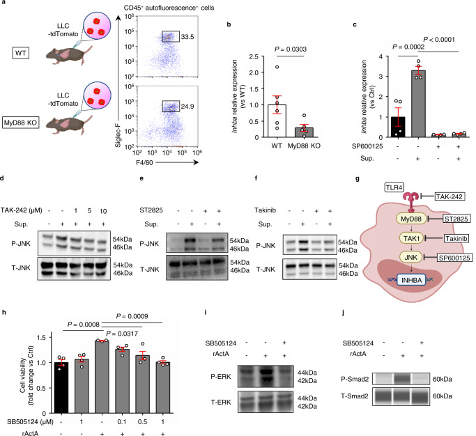
Fig. 4. INHBA expression in alveolar macrophages (AMs) is induced via MyD88-JNK dependent pathway.
a Flow cytometry plots of AMs isolated from C57BL/6 wild-type (WT) mice and MyD88 knockout (KO) mice after inoculation with lung cancer cells. b Inhba RT-PCR analysis of AM isolated from tumor-bearing wild-type mice and MyD88 knockout mice (n = 6 mice for wild-type, n = 5 mice for MyD88 KO). c Inhba RT-PCR analysis in the AM cell line AMJ2-C11 after incubation with or without LLC cell line supernatant (Sup.) and JNK inhibitor SP600125 (n = 4 per group). d–f Immunoblotting of JNK phosphorylation in AM cell line with or without stimulation of LLC cell line supernatant after incubation with each inhibitor (d; TAK-242, TLR4 inhibitor, e; ST2825, MyD88 inhibitor, f; takinib, TAK1 inhibitor). These images were representative of three independent experiments with similar results. g, Schematic representation of Inhba signaling in AM. h Viability of LLC cells treated with the ALK4 inhibitor SB505124 and recombinant activin A (rActA) was assessed using WST-1 assay (n = 4 per group except for the one with only rActA administration (n = 3)). i, j Immunoblotting of ERK (i) and Smad2 phosphorylation (j) in LLC cells stimulated with rActA after incubation with the ALK4 inhibitor SB505124. These images were representative of three independent experiments with similar results. Means ± s.e.m. for each group are shown. Symbols represent individual mice (b) and wells (c, h). Statistical significance was determined using unpaired two-tailed Mann–Whitney U-test (b) or one-way ANOVA with Bonferroni’s post hoc test (c, h).

