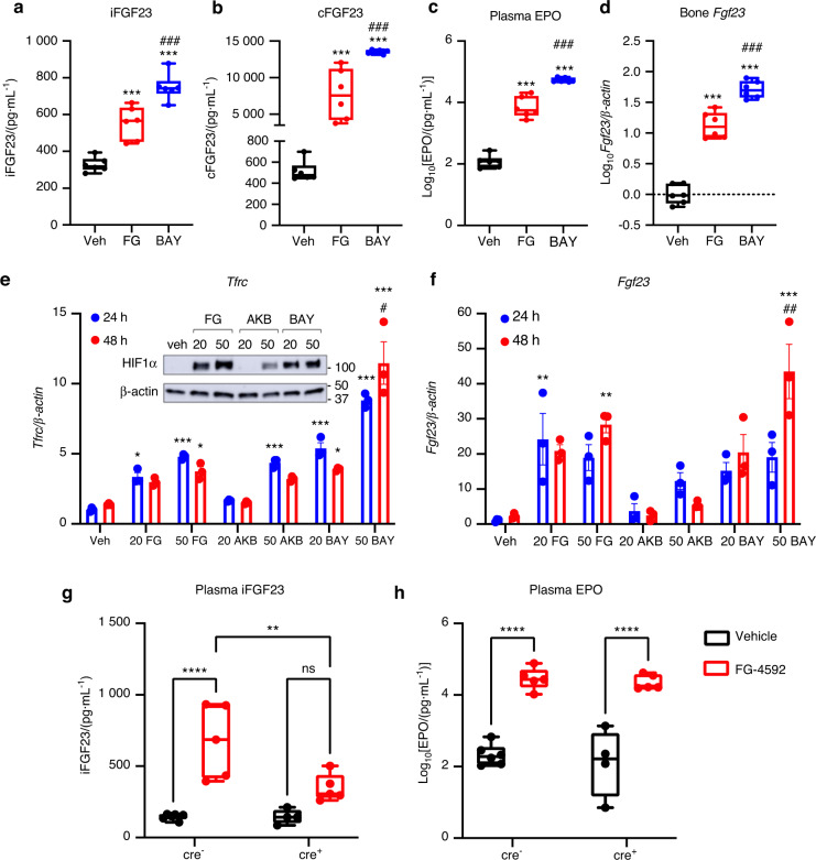
Fig. 1.
HIF-PHI induce FGF23 in WT mice and murine osteocyte-like cells. Wild type mice (females) were injected with vehicle (PEG300/DMSO) or 50 mg·kg−1 of either FG-4592 (‘FG’, Roxadustat; red) or BAY 85-3934 (‘BAY’, Molidustat; blue) every other day for 5 days, for a total of 3 injections. a Plasma intact FGF23 and b C-terminal/total FGF23 concentrations at 4 hours after final injection. c Plasma erythropoietin (EPO) concentrations and d cortical bone (flushed of marrow) Fgf23 mRNA expression at the end of the study (n = 6 mice per group; ***P < 0.001 versus vehicle, ###P < 0.001 versus FG treatment). e Transferrin receptor (Tfrc) and f Fgf23 mRNA expression in 2-week differentiated MPC2 cells treated for 24 h (blue) or 48 h (red) with 20 μmol·L−1 or 50 μmol·L−1 of the HIF-PHI FG-4592 (‘FG’; Roxadustat), AKB-6548 (‘AKB’; Vadadustat), or BAY 85-3934 (‘BAY’; Molidustat) (*P < 0.05, **P < 0.01, ***P < 0.001 versus vehicle; #P < 0.05, ##P < 0.01 24 h versus 48 h). (e, inset) HIF1α protein expression in MPC2 cells after 4 h of 20 μmol·L−1 or 50 μmol·L−1 HIF-PHI treatment. Flox-Fgf23/Dmp1-cre+ and cre− mice were injected with vehicle (black) or 70 mg·kg−1 FG-4592 (red) every other day for 5 days, for a total of 3 injections. Plasma concentrations of g intact FGF23 and h EPO were measured 4 hours after final injection (n = 4–6 mice per group; **P < 0.01, ****P < 0.000 1)

