Figure 1.
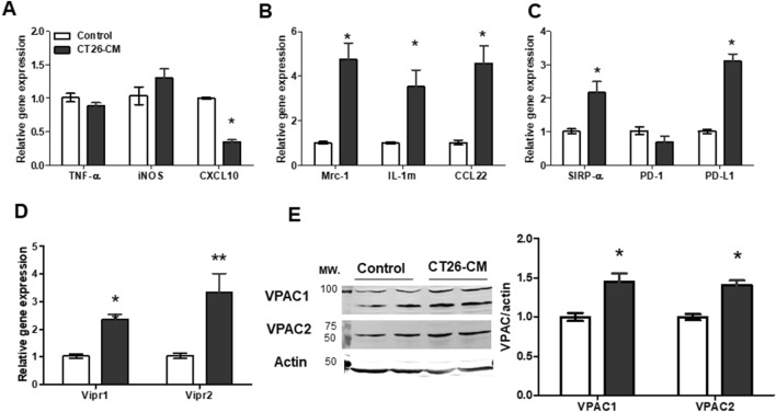
Conditioned medium from CT26 cells induced M2 macrophage polarization and VPAC expression in RAW264.7 cells. Real-time PCR analyses of M1 macrophage markers TNF-α, iNOS, and CXCL10 (A), M2 macrophage markers Mrc-1, IL-1rn, and CCL-22 (B), immune checkpoint markers SIRP-α, PD-1, and PD-L1 (C), and VIP receptors Vipr1 and Vipr2 (D) in RAW264.7 cells at day 4 after incubation with 20% conditioned medium derived from CT26 with DMEM complete medium (CT26-CM) or 20% RPMI medium with DMEM complete medium (control); n = 5–6/group. Representative western blotting images showing protein expression levels of VPAC1 and VPAC2 in RAW264.7 cells at day 4 after incubation with CT26-CM or control (E, left). Western blotting analysis of VPAC1 and VPAC2 protein expression in RAW264.7 cells at day 4 after incubation with control medium or CT26-CM (E, right); n = 5/group. The full-length blots of (E) are included in Supplemental Fig. S1. *p < 0.05 vs control, **p < 0.01 vs control.

