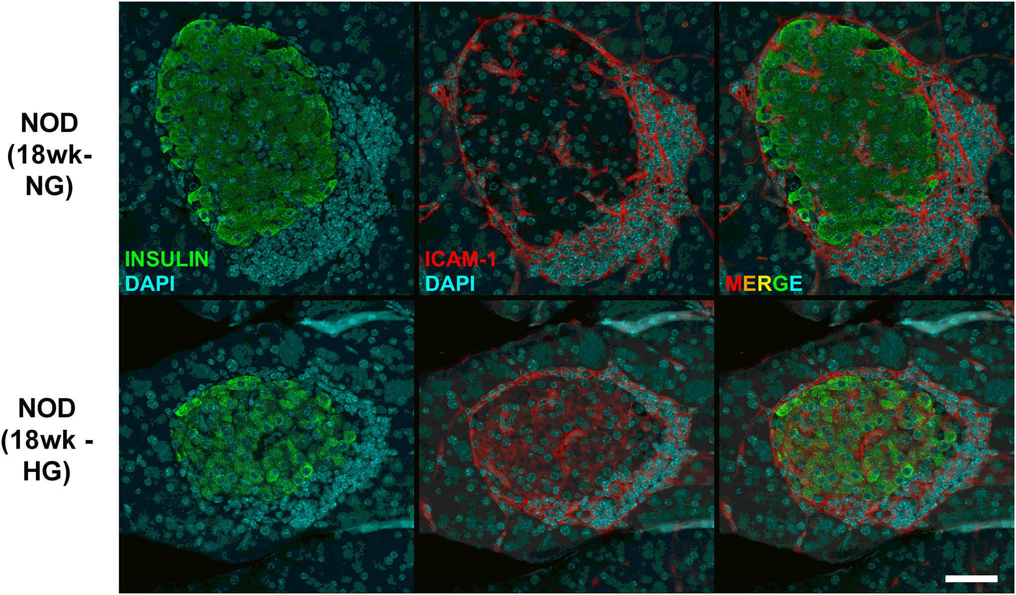
Figure 3. Hyperglyemic female NOD mice display ICAM-1 expression in pancreatic β-cells.

Pancreatic tissue from female NOD mice were collected at 18 weeks of age after two consecutive measurements of fed blood glucose values > 250 mg/dL (HG). Age matched normoglycemic controls (NG) were also collected at the same necropsy. Formalin-fixed paraffin-embedded pancreatic tissue was sectioned and stained using antibodies against insulin (shown in green) and ICAM-1 (shown in red) and imaged using a 40x objective. The scale bar represents 50 microns.
