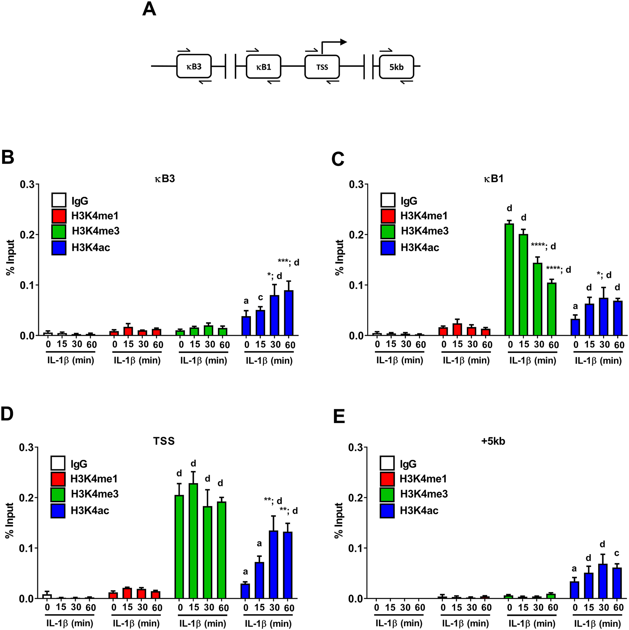
Figure 9. Histone chemical modifications associated with the Icam1 gene are regulated in response to IL-1β.

A. Schematic indicating the sites investigated for binding events using ChIP assays. B-E. 832/13 cells were untreated (0 min) or stimulated with 1 ng/mL IL-1β for the indicated times. ChIP assays were used to determine histone chemical modification (H3K4me1, H3K4me3, or H3K4ac) at the indicated sites. Rabbit IgG was used as a negative control. Data is represented as percent of input, with 3–4 replicates of each condition. Letters compare same IL-1β treatment time for IgG antibody; asterisks compare to 0 min time-point of respective antibody. *, p < 0.05; **, p < 0.01; ***, p < 0.001; ****, p < 0.0001. a p < 0.05, c p < 0.001, d p < 0.0001.
