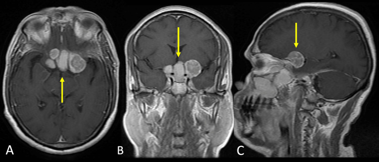
Figure 4. Magnetic resonance imaging with contrast images.
A. Axial T1WI contrast section showing multiple heterogeneously enhancing mass in the supra and parasellar region suggesting intracranial involvement of disease (yellow arrow).
B. Coronal T1WI contrast section showing multiple heterogeneously enhancing mass in the supra and parasellar region suggesting intracranial involvement of disease (yellow arrow).
C. Sagittal T1WI contrast section showing heterogeneously enhancing mass in the suprasellar region suggesting intracranial involvement of disease (yellow arrow).

