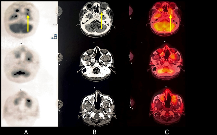
Figure 7. Positron emission tomography-computed tomography scan.
A. Positron emission tomography-computed tomography scan of the head reveals an ill-defined minimally enhancing mass lesion in the masticator space on the left side, showing low-grade diffuse fluorodeoxyglucose uptake with standardized uptake value max of 6.1 with obliteration of the retroantral fat on the left side. There is a superior extension of the mass reaching up to the apex of the left orbit (yellow arrow).
B. Contrast-enhanced computed tomography scan of the head reveals an ill-defined minimally enhancing mass lesion in the masticator space on the left side, showing low-grade diffuse fluorodeoxyglucose uptake with standardized uptake value max of 6.1 with obliteration of the retroantral fat on the left side. There is a superior extension of the mass reaching up to the apex of the left orbit (yellow arrow).
C. Positron emission tomography-computed tomography fusion scan of the head reveals an ill-defined minimally enhancing mass lesion in the masticator space on the left side, showing low-grade diffuse fluorodeoxyglucose uptake with standardized uptake value max of 6.1 with obliteration of the retroantral fat on the left side. There is a superior extension of the mass reaching up to the apex of the left orbit (yellow arrow).

