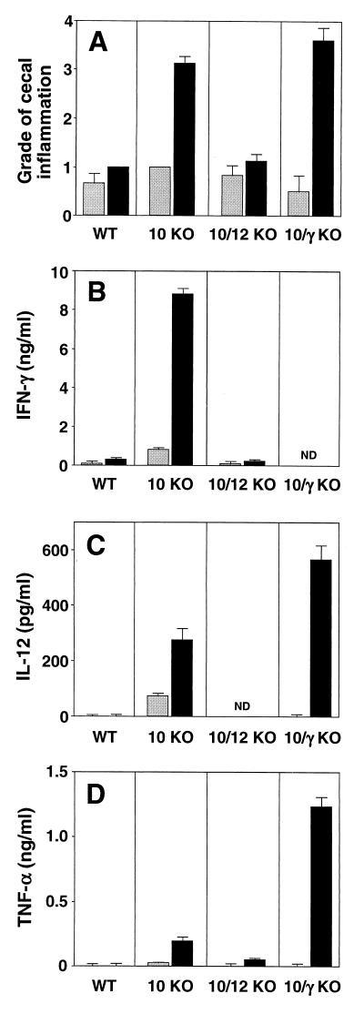
FIG. 9.
IL-10/IFN-γ KO mice develop colitis following inoculation with H. hepaticus. WT, IL-10 KO, IL-10/IL-12 KO, and IL-10/IFN-γ KO animals were infected i.g. with H. hepaticus (black bars), and intestinal pathology and cytokine responses were analyzed 4 weeks later. Gray bars represent uninfected controls. (A) Pathology in the cecum. Bars represent mean histology scores ± SE of 3 to 5 mice/group, with the following median values for infected mice: WT = 1 (all mice scored as 1, n = 3), IL-10 KO = 3 (range, 3 to 3.5, n = 4), IL-10/IL-12 KO = 1 (range, 1 to 1.5, n = 4), and IL-10/IFN-γ KO = 4 (range, 3 to 4, n = 5). Similar results were observed for colon, although scores were lower (not shown). (B to D) IFN-γ (B), IL-12 (C), and TNF-α (D) levels detected in 72-h supernatants from MLN cell cultures stimulated with SHelAg. Bars represent means ± SD of duplicate ELISA values from MLN cells pooled from 3 to 5 mice/group. The data shown are from one representative experiment of two performed. ND, not detected. No IFN-γ, IL-12, or TNF-α was detected from cells cultured in medium alone, and no IL-4 was detected in supernatants from SHelAg-stimulated cultures.

