Figure 1.

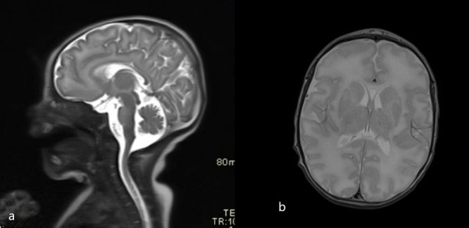
Initial brain MRI scans (T2 sagittal and axial plane). (A) Normal thickness of pons and midbrain. (A, B) Higher T2 signal of the frontal white matter.

Initial brain MRI scans (T2 sagittal and axial plane). (A) Normal thickness of pons and midbrain. (A, B) Higher T2 signal of the frontal white matter.