Figure 3.

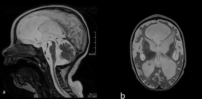
(A) Follow-up brain MRI scans (T2 sagittal and axial plane) after 1 year: severe atrophy of midbrain compared with initial MRI. (B) Severe reduction of brain volume and abnormal signal of the white matter—leucoencephalopathy.

(A) Follow-up brain MRI scans (T2 sagittal and axial plane) after 1 year: severe atrophy of midbrain compared with initial MRI. (B) Severe reduction of brain volume and abnormal signal of the white matter—leucoencephalopathy.