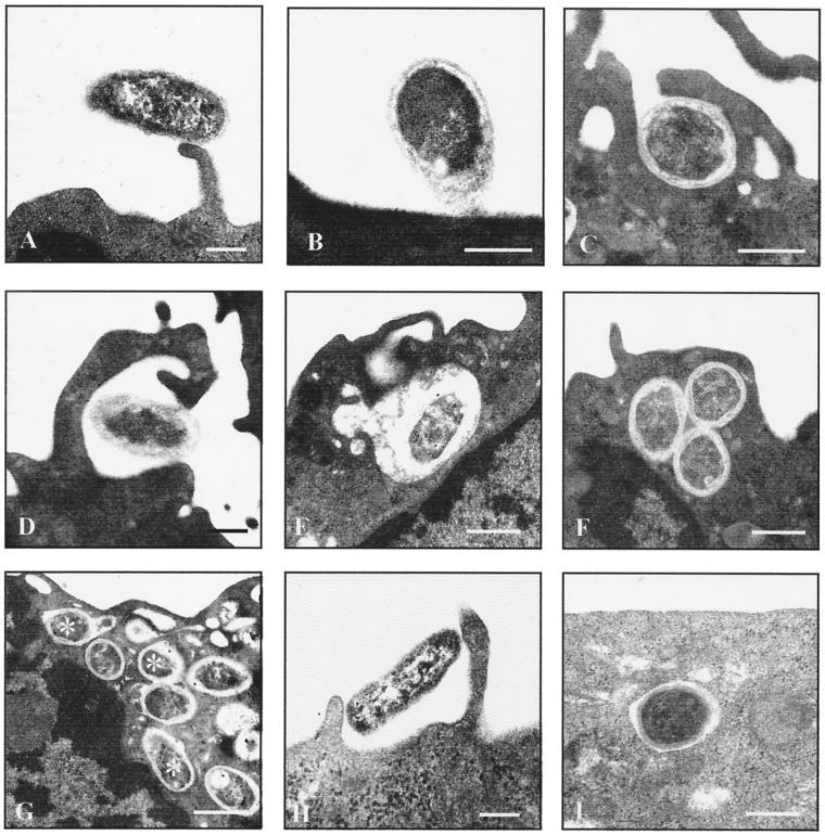
FIG. 1.
Ultrastructural observations on the phagocytosis of Brucella by human monocytes and epithelioid cells. Freshly isolated monocytes (A to G) or epithelioid cells (H and I) were challenged with nonopsonized B. suis 1330 for 15 min and chased for up to 24 h. Bars, 0.2 μm. Brucellae attached to various parts of the plasma membrane, such as cell surface projections (A) or the plain cell surface (B). The emerging phagocytic cups which formed within the first minutes had either continuous (C) or focal (D) contact with the bacteria. The resulting phagosomes either were spacious (E) or had tightly apposed walls (F). Both types of phagosomes showed fusion with small intracellular vesicles, with the fusion events for the latter type being more numerous after longer chasing periods (G) (asterisks indicate phagosome-lysosome fusion; chase was for 2 h). Uptake of Brucella by HeLa or CHO cells also took place via the usual phagocytic mechanisms (H) (HeLa cells) and resulted in membrane-bound compartments which predominantly had tightly apposed walls (I) (CHO cells).

