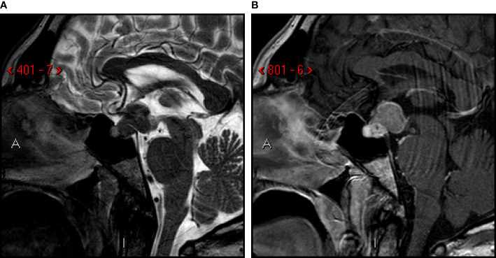
Figure 1.
First MRI-scan. Sagittal T2-weighted (A) and post-contrast T1-weighted (B) images. Sellar and suprasellar mass involving posterior lobe and infundibulum of the pituitary gland, with low signal on T2-images. Compression of mammillary body, optic chiasm and tracts, third ventricle (infundibular recess).

