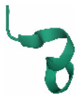
Table 7.
The amino acid sequence and identified methods for Aβ peptide.
| Types | Molecularformula | Molecular | Amino acid sequence | 3D Structure | Structure-neurotoxicity | Characteristic | Identified methods | Reference | |||||||||||||||||
|---|---|---|---|---|---|---|---|---|---|---|---|---|---|---|---|---|---|---|---|---|---|---|---|---|---|
| I31-V40 | CHC | I31-V40 C | alpha-helixconformation | β-sheet conformation | active region of Aβ | proteolytic cleavage | CHC | G37-G38 | C-terminal amino acidsIle41-Ala42 | β-strand structure involving Ala-2–Phe-4 | CD | NMR | AFM | ThT | EFM | SKM | cryo-EM | IEM | Other | ||||||
| Aβ25 − 35 | C45H81N13O14S | 1060.27 | H-Gly25-Ser-Asn-Lys-Gly-Ala-Ile-Ile-Gly-Leu-Met35-OH |
 PDB (1QWP) PDB (1QWP) |
√ | √ | √ | √ | √ | √ | √ | √ | √ | Yankner et al., 1990; Terzi et al., 1994; Sato et al., 1995; Kubo et al., 2002; Naldi et al., 2012; Chen et al., 2017 | |||||||||||
| Aβ1 − 42 | C203H311N55O60S | 4514.1 | H-Asp-Ala-Glu-Phe-Arg-His-Asp-Ser-Gly-Tyr-Glu-Val-His-His-Gln-Lys-Leu-Val-Phe-Phe-Ala-Glu-Asp-Val-Gly-Ser-Asn-Lys-Gly-Ala-Ile-Ile-Gly-Leu-Met-Val-Gly-Gly-Val-Val-Ile-Ala-OH |
 PDB (1IYT) |
√ | √ | √ | √ | √ | √ | √ | √ | √ | √ | √ | colorimetric, fluorometric methods, differential interference contrast optics, laser scanning confocal immunofluorescence | Naiki and Gejyo, 1999; Anderson et al., 2004; Urbanc et al., 2004, 2010; Inouye and Kirschner, 2005; Bartolini et al., 2007; Middleton, 2007; Ahmed et al., 2010; Chen et al., 2017 | ||||||||
| Aβ1 − 40 | C194H295N53O58S1 | 4329.9 | H-Asp-Ala-Glu-Phe-Gly-His-Asp-Ser-Gly-Phe-Glu-Val-Arg-His-Gln-Lys-Leu-Val-Phe-Phe-Ala-Glu-Asp-Val-Gly-Ser-Asn-Lys-Gly-Ala-Ile-Ile-Gly-Leu-Met-Val-Gly-Gly-Val-Val-OH |
 PDB (1AML) |
√ | √ | √ | √ | √ | √ | √ | √ | Urbanc et al., 2004, 2010; Williams et al., 2005; Sachse et al., 2006; Meinhardt et al., 2009; Bertini et al., 2011; Naldi et al., 2012; Chen et al., 2017 | ||||||||||||
The table mainly describes the molecular and molecular formula, amino acid sequence, structure and identified methods in Aβ peptide. To present the results in a systematic manner, the peptide is segmented as follows: (i) Asp-1–Lys-16 is the N-terminal region; (ii) Leu-17–Ala-21 is the central hydrophobic cluster (CHC); (iii) Glu-22–Gly-29 is the turn A (TRA) region; (iv) Ala-30–Met-35 is the mid-hydrophobic region (MHR); (v) Val-36–Val-39 is the turn B (TRB) region; and (vi) Val-40 or Val-40–Ala-42 is the C-terminal region (CTR).
EFM, electrostatic force microscopy; AFM, atomic force microscopy; SKM, scanning Kelvin microscopy; cryo-EM, cryo-electron.
