FIG. 3.
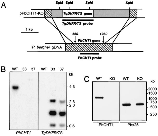
Targeted disruption of PbCHT1 and molecular analyses. (A) Schematic diagram of the targeting strategy. Indicated is the transfection vector pPbCHT1-KO containing the T. gondii DHFR/TS gene cassette (white box), P. berghei DHFR flanking sequences (gray boxes), and PbCHT1-specific sequences (hatched boxes). The double homologous recombination crossover sites (crossed lines), the integration sites (arrows with nucleotide positions), the SphI restriction sites, and the probes used in Southern blot analysis (thick lines) are shown. gDNA, genomic DNA. (B) Southern blot analysis of SphI-digested genomic DNA from WT and PbCHT1-KO parasites using probes corresponding to PbCHT1 (left panel) and to the DHFR/TS cassette (right panel). (C) RT-PCR analysis of total RNA derived from ookinete-enriched midgut stages of WT (left lanes) and PbCHT1-KO (right lanes) parasites. Amplicons corresponding to PbCHT1(∼1,100 bp) and Pbs25 (∼600 bp) are shown.

