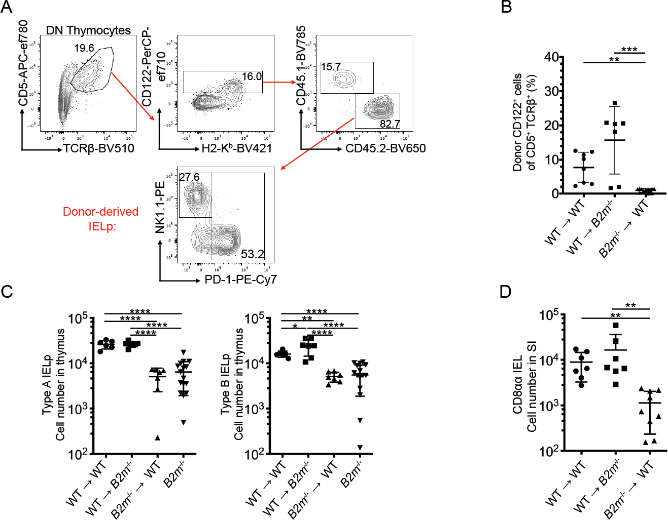
Figure 1. Positive selection of IELp depends on MHC class I expressed on hematopoietic APC.

(A) Representative flow cytometry plots of DN thymocytes in bone marrow chimeras (WT → WT shown). Full gating for IELp shown in Supp. Fig. 1. Mature IELp were defined by CD122 expression alone due to the lack of H2-Kb expression in mice with β2m deficient bone marrow. Donor and host cells were differentiated using congenic markers, CD45.1 and CD45.2. Numbers adjacent to the outlined areas indicate the percentage of cells in each. (B) Quantified percentage of donor-derived IELp after WT or B2m−/− bone marrow transfer into lethally irradiated recipients indicated. (C) Absolute number of PD-1+ Type A IELp (left) and NK1.1+ Type B IELp (right) after WT or B2m−/− bone marrow transfer into lethally irradiated recipients indicated. (D) Absolute number of small intestine CD8αα IEL in the indicated bone marrow chimeras. Each symbol in (B-D) represents an individual mouse [n=8 for WT → WT in (B-C), n=7 in (D); n=7 for WT→B2m−/− in (B-D); n=7 for B2m−/− →WT in (B-C), n=9 in (D); n=16 for B2m−/− in (C)]. Data are pooled from at least three independent experiments. Error bars show mean ± SD. *p≤0.05, **p≤0.01, ***p≤0.001, ****p ≤ 0.0001, ANOVA with multiple comparisons.
