
Figure 4. Conditional deletion of β2m in conventional DC drive more cells toward the Type A IELp fate.
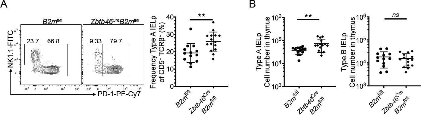
(A) Expression of NK1.1 (Type B IELp) and PD-1 (Type A IELp) on CD122+ H2-Kb+ mature thymic IELp in Zbtb46Cre (zDCCre)-B2mfl/fl and littermate control mice (left, representative data are shown). Numbers adjacent to the outlined areas indicate the percentage of cells in each. Quantified percentage of PD-1+ Type A IELp in zDCCre-B2mfl/fl and B2mfl/fl mice among signaled CD5+ TCRβ+ DN thymocytes (right). (B) Absolute numbers of PD-1+ Type A IELp and NK1.1+ Type B IELp in zDCCre-B2mfl/fl and B2mfl/fl mice. Each symbol in (A & B) represents an individual mouse (n=13 for B2mfl/fl and n=15 for Zbtb46Cre-B2mfl/fl). Data are pooled from 7 independent experiments. Error bars show mean ± SD. **p≤0.01, unpaired two-tailed t test. All data was measured by flow cytometry.