. 2023 Jan 10;306(2):e222462. doi: 10.1148/radiol.222462

Figure 10:

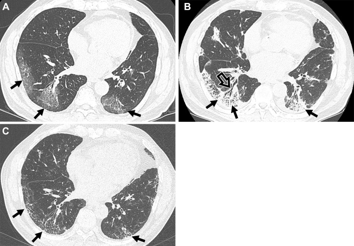
Serial images in a 72-year-old man with COVID-19 pneumonia. (A) Transverse nonenhanced CT scan (lung window) obtained at inferior pulmonary vein level 1 week after SARS-CoV-2 infection shows lower lobe–predominant patchy areas of ground-glass opacity (GGO) in bilateral lungs (arrows). (B) Transverse nonenhanced CT scan (lung window) obtained at inferior pulmonary vein level 1 month after SARS-CoV-2 infection shows lower lobe–predominant patchy areas of consolidation (solid arrows) in bilateral lungs. Note bronchial dilatation (open arrow) within the consolidation. (C) Transverse nonenhanced CT scan (lung window) obtained 6 months after infection demonstrates residual reticulations (arrows) and GGOs.

Serial images in a 72-year-old man with COVID-19 pneumonia. (A) Transverse nonenhanced CT scan (lung window) obtained at inferior pulmonary vein level 1 week after SARS-CoV-2 infection shows lower lobe–predominant patchy areas of ground-glass opacity (GGO) in bilateral lungs (arrows). (B) Transverse nonenhanced CT scan (lung window) obtained at inferior pulmonary vein level 1 month after SARS-CoV-2 infection shows lower lobe–predominant patchy areas of consolidation (solid arrows) in bilateral lungs. Note bronchial dilatation (open arrow) within the consolidation. (C) Transverse nonenhanced CT scan (lung window) obtained 6 months after infection demonstrates residual reticulations (arrows) and GGOs.