. 2023 Jan 10;306(2):e222462. doi: 10.1148/radiol.222462

Figure 11:

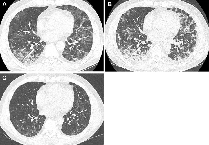
Serial images in a 55-year-old man with COVID-19 pneumonia. (A) Transverse nonenhanced CT scan (lung window) obtained at levels of basal segmental bronchi 4 weeks after SARS-CoV-2 infection shows extensive and patchy areas of mixed ground-glass opacity (GGO) and consolidation in bilateral lungs. (B) Transverse nonenhanced CT scan (lung window) obtained 5 weeks after infection demonstrates increased density of GGO lesions with consolidation. (C) Transverse nonenhanced CT scan (lung window) obtained 9 months after infection demonstrates residual faint GGO and reticulations.

Serial images in a 55-year-old man with COVID-19 pneumonia. (A) Transverse nonenhanced CT scan (lung window) obtained at levels of basal segmental bronchi 4 weeks after SARS-CoV-2 infection shows extensive and patchy areas of mixed ground-glass opacity (GGO) and consolidation in bilateral lungs. (B) Transverse nonenhanced CT scan (lung window) obtained 5 weeks after infection demonstrates increased density of GGO lesions with consolidation. (C) Transverse nonenhanced CT scan (lung window) obtained 9 months after infection demonstrates residual faint GGO and reticulations.