. 2023 Jan 10;306(2):e222462. doi: 10.1148/radiol.222462

Figure 12:

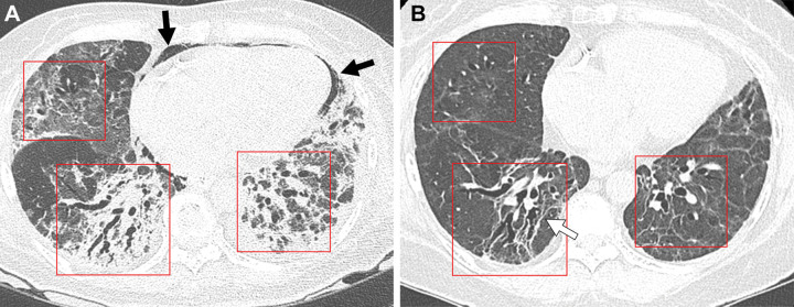
Serial images in a 64-year-old woman with COVID-19 pneumonia. (A) Transverse nonenhanced CT scan (lung window) obtained at ventricular level 1 month after SARS-CoV-2 infection shows lower lobe–predominant patchy and wide areas of mixed consolidation and ground-glass opacity (GGO) in bilateral lungs. Note areas of bronchial dilatation (boxes) within parenchymal lesions. Pneumomediastinum (arrows) is also present anteriorly. (B) Transverse nonenhanced CT scan (lung window) obtained 12 months after infection demonstrates dilated bronchi (boxes) within remaining GGO lesions. Linear parenchymal bands (arrow) are also noted.

Serial images in a 64-year-old woman with COVID-19 pneumonia. (A) Transverse nonenhanced CT scan (lung window) obtained at ventricular level 1 month after SARS-CoV-2 infection shows lower lobe–predominant patchy and wide areas of mixed consolidation and ground-glass opacity (GGO) in bilateral lungs. Note areas of bronchial dilatation (boxes) within parenchymal lesions. Pneumomediastinum (arrows) is also present anteriorly. (B) Transverse nonenhanced CT scan (lung window) obtained 12 months after infection demonstrates dilated bronchi (boxes) within remaining GGO lesions. Linear parenchymal bands (arrow) are also noted.