. 2023 Jan 10;306(2):e222462. doi: 10.1148/radiol.222462

Figure 8:

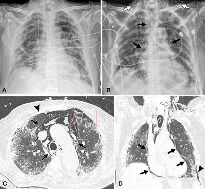
Images of COVID-19 pneumonia with oxygen toxicity and barotrauma at the time of Delta variant–dominant period in a 71-year-old unvaccinated man with diabetes. (A) Chest radiograph obtained 10 days after positive nucleic acid amplification test for COVID-19 shows extensive parenchymal opacity involving bilateral lungs. He was transferred to the intensive care unit due to worsening of hypoxemia and was given mechanical ventilation with prolonged oxygen supply. (B) Follow-up chest radiograph obtained 25 days after initial diagnosis of COVID-19 demonstrates identifiable pneumomediastinum (black arrows) and subcutaneous emphysema (white arrows) with parenchymal opacities. (C) Transverse and (D) coronal reformatted images of nonenhanced CT scans depict mixed ground-glass opacity, consolidation, and reticulation in the peripheral areas of both lungs associated with interstitial emphysema (small arrows in C) causing Macklin effect, pneumothorax (arrowhead), pneumomediastinum (black arrows) and subcutaneous emphysema (white arrows in D). Note air cysts in the anterior left lung (box in C). One month after receiving mechanical ventilation with corticosteroid treatment, the patient recovered and was discharged.

Images of COVID-19 pneumonia with oxygen toxicity and barotrauma at the time of Delta variant–dominant period in a 71-year-old unvaccinated man with diabetes. (A) Chest radiograph obtained 10 days after positive nucleic acid amplification test for COVID-19 shows extensive parenchymal opacity involving bilateral lungs. He was transferred to the intensive care unit due to worsening of hypoxemia and was given mechanical ventilation with prolonged oxygen supply. (B) Follow-up chest radiograph obtained 25 days after initial diagnosis of COVID-19 demonstrates identifiable pneumomediastinum (black arrows) and subcutaneous emphysema (white arrows) with parenchymal opacities. (C) Transverse and (D) coronal reformatted images of nonenhanced CT scans depict mixed ground-glass opacity, consolidation, and reticulation in the peripheral areas of both lungs associated with interstitial emphysema (small arrows in C) causing Macklin effect, pneumothorax (arrowhead), pneumomediastinum (black arrows) and subcutaneous emphysema (white arrows in D). Note air cysts in the anterior left lung (box in C). One month after receiving mechanical ventilation with corticosteroid treatment, the patient recovered and was discharged.