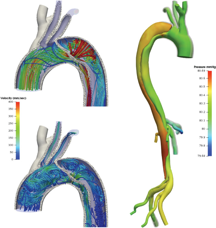
FIG. 2.
FSI model of aortic dissection with vessel wall prestress and external tissue support. Streamlines (left) in systole (top) and diastole bottom and pressure (right). True and false lumen are clearly visible with streamlines entering the false lumen at the focal entry tear. Adapted from Ref. 27.

