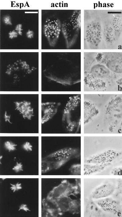
FIG. 4.
EspA expression and A/E lesion formation on HEp-2 cells produced by EspD coiled-coil mutants. EspA fluorescence (column 1), FAS test actin fluorescence (column 2) and corresponding phase-contrast micrographs (column 3) of wild-type EPEC strain E2348/69 (a), espD deletion mutant strain UMD870 (b), cloned EspD strain UMD870(pLCL123) (c), double EspD coiled-coil mutant strain UMD870(pICC73) (d), and triple EspD coiled-coil mutant strain UMD870(pICC74) (e). The coiled-coil mutants expressed EspA filaments (d, e), but whereas the double mutant produced a positive FAS reaction (actin accumulation at sites of bacterial attachment) (d), the triple mutant produced a barely detectable FAS reaction (e). Note that the espD deletion mutant produced barely detectable EspA filaments (b) and that EspA filaments expressed from strains harboring plasmid pLCL123 (c to e) are of the same length. Bars, 5 μm (column 1) and 20 μm (columns 2 and 3).

