Fig. 1.
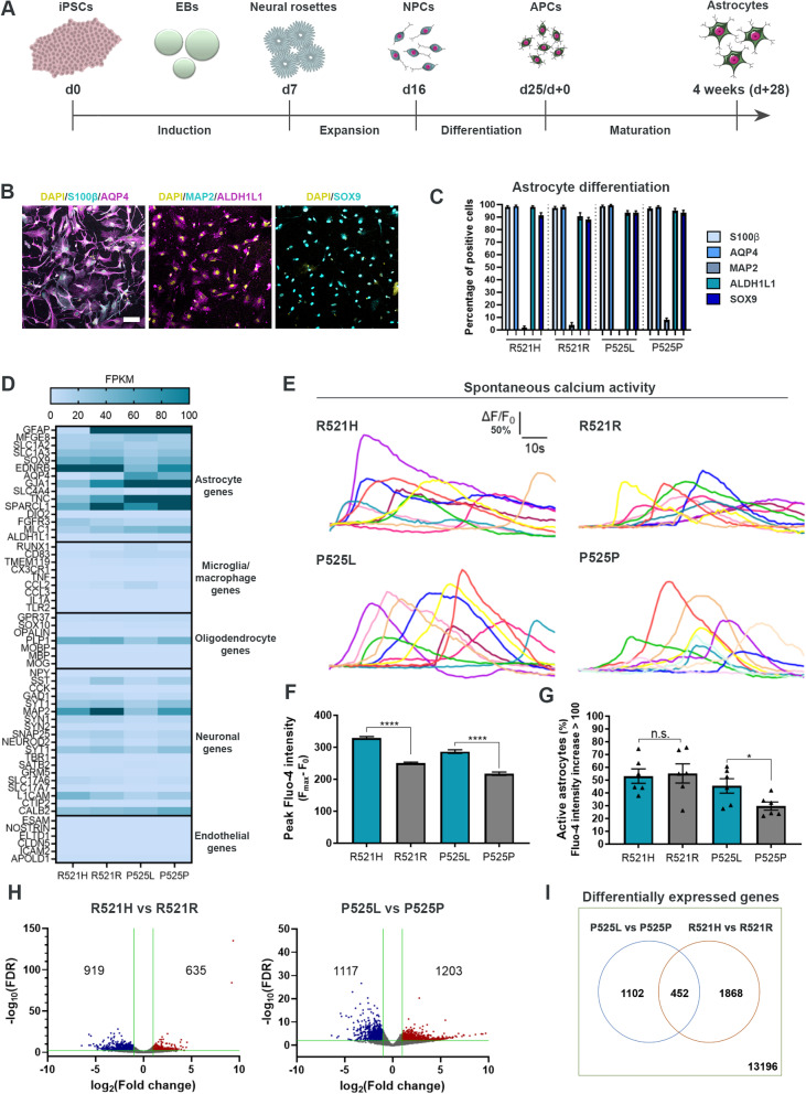
Generation and characterisation of functional hiPSC-derived astrocytes. A Overview of the astrocyte differentiation protocol. hiPSCs were dissociated at day 0 (d0) and cultured for the induction phase as embryoid bodies (EBs) before they were plated for neural rosette formation at day 7 (d7). The neural rosettes underwent expansion to generate neural progenitor cells (NPCs) until day 16 (d16). NPCs were then differentiated into astrocyte progenitor cells (APCs, d25/d+0) after which the APCs were matured into astrocytes for an additional 4 weeks (d + 28). B Representative confocal images of astrocytes at week 4 (d + 28) of maturation stained with astrocytic markers S100β, AQP4, ALDH1L1 and SOX9 in addition to neuronal marker MAP2. Nuclei stained with DAPI. Scale bar: 75 μm. C Quantification of the number of cells positive for astrocyte and neuronal markers. Mean ± s.e.m. of 3 biological replicates (n = 15 images). D Heatmap of the expression of cell type-specific genes in 4 weeks mature astrocytes from RNAseq experiment performed in 3 biological replicates. Dark blue cells: FPKM > 100. E Functional assessment of spontaneous calcium transients in 6 weeks mature astrocytes loaded with the Fluo-4 dye. Y-axis range: ΔF/F0 = 50%. F Quantification of peak Fluo-4 intensity. Mann–Whitney test. ****p < 0.0001. G Percentage of active astrocytes. Inactive astrocyte cut-off: Fluo-4 intensity increase < 100. Unpaired t test. *p < 0.05. H Volcano-plots of up- (red) and down- (blue) regulated genes in week 4 mature astrocytes. Green lines indicate thresholds: log2FC < -1.0 with –log10(FDR) > 2.0 were considered downregulated, and log2FC > 1.0 with –log10(FDR) > 2.0 were considered upregulated. I Comparison of differentially expressed genes between mutation sets. Graphs in panels (F and G) show mean ± s.e.m. of 3 biological replicates with each 2–6 technical replicates. Cell illustrations are modified from Smart Servier Medical Art licensed under a Creative Commons Attribution 3.0 Unported License (https://creativecommons.org/licenses/by/3.0/). See also Suppl. Figure 1–3, Additional file 2, as well as Additional files 3, 4, 5 and 6

