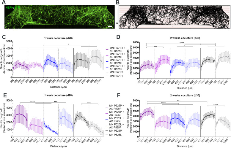
Fig. 5.
P525L FUS-ALS astrocytes impair motor neuron neurite outgrowth. A Example of a tile scan confocal image of motor neuron (MN) neurite (NEFH/SYP) outgrowth in the myotube compartment after 1 week (d28) of coculture with astrocytes (AC). Arrows (right) depict the neurite growth direction upon exit of the microgrooves. Scale bar: 300 μm. B Mask of tile scans with intersection lines at every 50 μm starting from the exit of the microgrooves. C-F Neurite outgrowth quantifications of the number of pixel intersections after 1 week (panel C + E) and 2 weeks (panel D + F) of coculture between motor neurons, myotubes and astrocytes. Graphs in (panel C-F) show mean ± s.e.m. of 3 biological replicates. Overall comparisons between genotypes were performed with two-way ANOVA with Tukey’s multiple comparisons test. *p < 0.05, **p < 0.01, ***p < 0.001 and ****p < 0.0001

