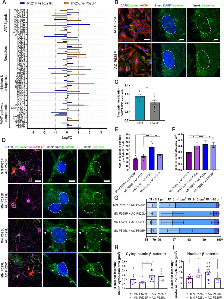
Fig. 7.
FUS-ALS astrocytes influence motor neurons through the WNT/β-catenin pathway. A RNAseq differential gene expression of WNT/β-catenin pathway components in 4-week mature astrocytes. B Representative confocal images of β-catenin expression in 4-week mature P525L and P525P astrocytes (AQP4). Inset shows a magnification of astrocytes with β-catenin localisation. Nuclei stained with DAPI. Scale bar: 50 μm. Inset scale bar: 5 μm. C Quantification of β-catenin localisation presented as the nuclei/cytoplasmic ratio. Data from 3 biological replicates (n = 30 images). Unpaired t test. **p < 0.01. D Representative confocal images of β-catenin expression in motor neurons (Tubulin), which have been cocultured with astrocytes (GFAP) for 48 h. Inset shows a magnification of motor neurons with β-catenin localisation. Arrowheads show examples of β-catenin accumulation. Nuclei (DAPI) are circled with a white dashed line. Scale bar: 50 μm. Inset scale bar: 5 μm. E Quantification of number of β-catenin accumulations per Tubulin+ motor neuron. F Quantification of individual β-catenin accumulation size in Tubulin+ motor neuron. G Percentage distribution of different size ranges of β-catenin accumulation in Tubulin+ motor neuron. H Quantification of cytoplasmic β-catenin expression per Tubulin+ motor neuron cytoplasmic area (μm2). I Quantification of nuclear β-catenin expression per Tubulin+ motor neuron nuclear area (μm.2). Panel (E-I) show mean ± s.e.m. of 3 biological replicates (n = 30 images). Panel (E and I): One-way ANOVA with Tukey’s multiple comparisons test. Panel (F and H): Kruskal–Wallis test with Dunn’s multiple comparison test. *p < 0.05, **p < 0.01, ***p < 0.001 and ****p < 0.0001

