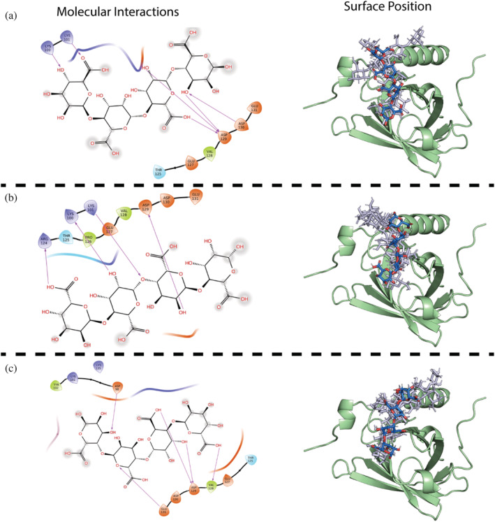
FIGURE 7.

Orientation of AOSs docked onto β‐LgA (green) binding site 4 (Lys101). Right hand side shows surface position of (a) M4 best XP GScore (blue), top 2–5 XP GScore (gray), (b) MG4 best XP GScore (blue), top 2–5 XP GScore (gray), (c) G4 best XP GScore (blue), top 2–5 XP GScore (gray). To the left are shown interaction schemes for the best XP GScore of OH, carboxyl and glycosidic oxygen groups in individual AOSs to residues at binding site 4. Acidic side chains are colored orange, basic side chains blue, hydrophobic side chains green and polar side chains cyan. Arrows mark side chain AOSs interactions
