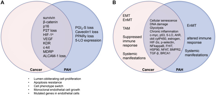
Fig. 1.
Venn diagrams showing the state of knowledge in 2008 (A) and in 2019 (B). The overlapping areas represent proteins, which are expressed both in cancers and in pulmonary arterial hypertension (PAH) lung tissue. ALCAM-1, activated leukocyte cell adhesion molecule; EMT, epithelial mesenchymal transition; EnMT, endothelial mesenchymal transition; HIF-1α, hypoxia-inducible factor-1α; KDR, VEGF receptor 2; 5-LO, 5-lipoxygenase; MDRP, multiple drug resistance protein; PGI2, prostacyclin; PPARγ, peroxisome proliferator-activated receptor-γ; TAM, tumor-associated macrophages. The overlap area in B represents protein expression data and mechanistic aspects of the “quasi-malignancy” concept like “glycolysis” and “DNA damage” that have been added to the literature since 2008. For more details, see Table 1.

