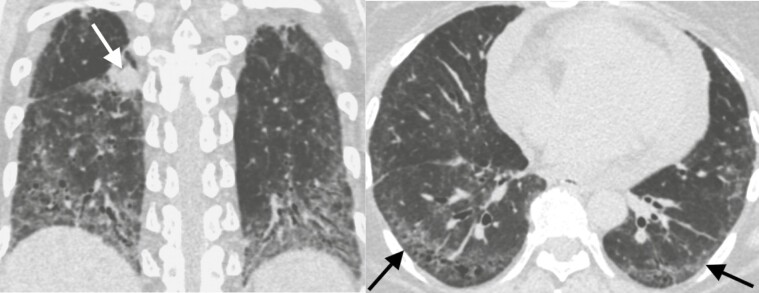
Figure 2.
Forty-four-year-old woman with systemic sclerosis-associated interstitial lung disease and an enlarging right lower lobe solid nodule corresponding to a biopsy proven non-small cell lung cancer. Left. Coronal CT image demonstrates the lung cancer (arrow) and lower lung predominant fibrosis marked by volume loss, ground-glass opacities, and traction bronchiectasis. Right. Axial CT image of the lung bases demonstrates peripheral predominant ground glass, reticular markings, and traction bronchiectasis (arrows) most consistent with a non-specific interstitial pneumonitis pattern.

