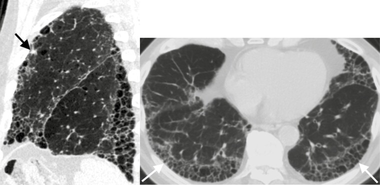
Figure 3.
Eighty-two-year-old man with idiopathic pulmonary fibrosis and an indeterminate subpleural solid left upper lobe nodule. Left: Sagittal CT image demonstrates the nodule (arrow) and honeycombing with an apical basilar gradient consistent with a usual interstitial pneumonia pattern. Right: Axial CT image of the lung bases demonstrates stacked cystic structures (arrows) consistent with honeycombing.

