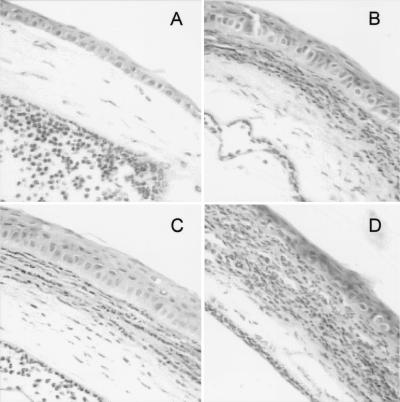
FIG. 1.
Histopathology of corneas from IL-6 gko and wild-type mice 12 h after challenge with P. aeruginosa. All sections are stained with hematoxylin and eosin. Magnification of all sections, ×176. (A) Cornea of IL-6 gko mouse 12 h after challenge with P. aeruginosa after subconjunctival injection of PBS. The central cornea contains only the occasional inflammatory cell. There are numerous PMN in the anterior chamber. (B) Cornea of wild-type mouse 12 h after challenge with P. aeruginosa after subconjunctival injection of PBS. The cornea contains infiltrating inflammatory cells, predominantly PMN. (C) Cornea of IL-6 gko mouse 12 h after challenge with P. aeruginosa after subconjunctival injection of IL-6. The central cornea contains inflammatory cells, predominantly PMN. There are numerous PMN in the anterior chamber. (D) Cornea of wild-type mouse 12 h after challenge with P. aeruginosa after subconjunctival injection of IL-6. The infiltrating inflammatory cells in the cornea are more numerous than in panel B.

