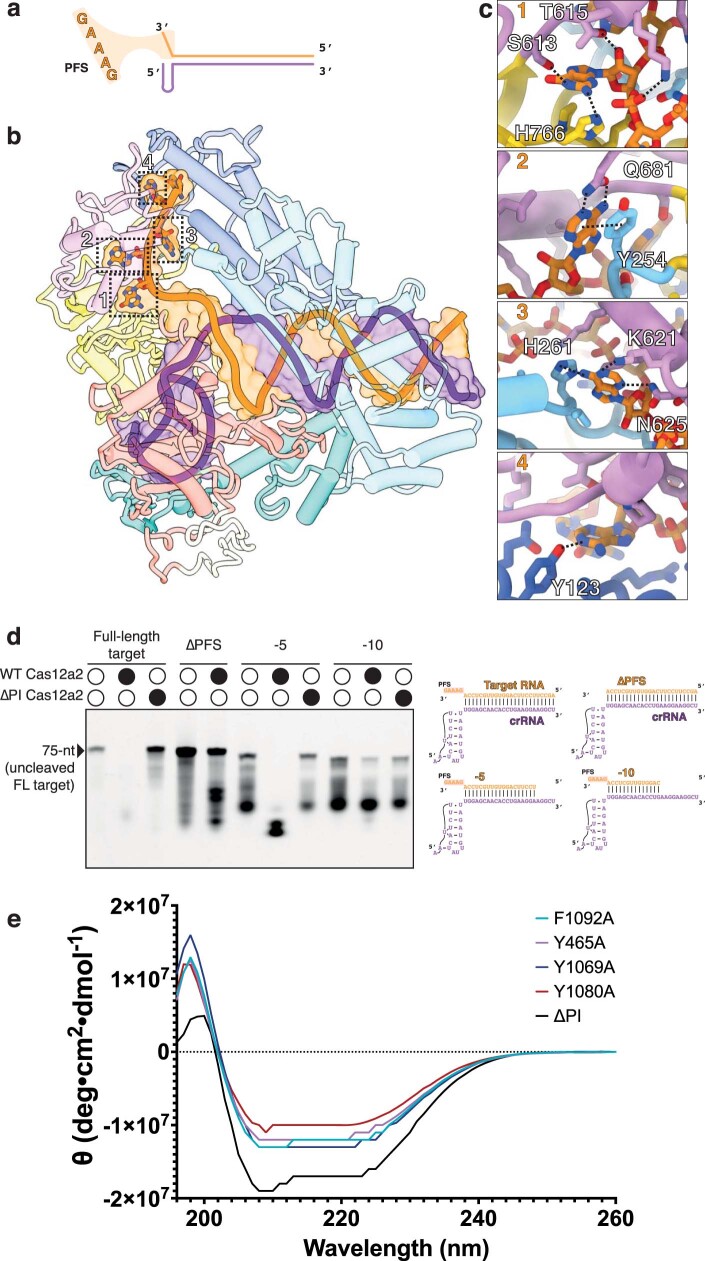
Extended Data Fig. 4. Mechanism of protospacer-flanking sequence (PFS) recognition by Cas12a2.
a, Schematic representation of PFS (GAAAG) located at the 3′ end of the RNA target RNA. b, Positions of the PFS within the Cas12a2 active ternary complex. c, Zoom-in of interactions between Cas12a2 and the first four bases of PFS. d, Activation of Cas12a2 or Cas12a2 ΔPI cleavage by truncated target RNAs. Representative of three independent experiments with similar results. e, Circular Dichroism (CD) spectroscopy of Cas12a2 mutants, including ∆PI truncation. All mutants are properly folded. For gel source data, see Supplementary Fig. 1.

