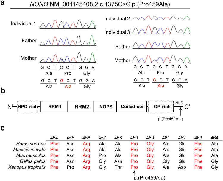
Figure 1.
Genetic analysis of NONO variant. (a) Electropherograms show that Individuals 1–3 had a hemizygous NONO variant inherited from their mother. The altered nucleotide and amino acid are shown in red. (b) Schematic presentation of NONO domains (solid line) and regions (dashed line), and the identified variant (arrow). NLS, nuclear localization signal; NOPS, NonA/paraspeckle; RRM, RNA recognition motif. (c) Evolutionary conservation of NONO protein and its orthologs within vertebrates. Conserved amino acids are shown in red.

